
Services
Imaging
Clearly focused on you
At Adventist Health Sonora, our advanced imaging technology gives your physician new clarity to better diagnose conditions and provide precision treatment. We believe your diagnostic imaging should be a calm, welcoming, and comfortable experience. That's why your exam will be conducted by well-trained staff focused on helping you every step of the way and ensuring your comfort and safety.
Comprehensive imaging services
We offer a full range of the latest imaging technologies for diagnosing and evaluating a broad range of conditions. We also have a staff of technicians and radiologists who are highly trained in using our technologies, reporting your results, and consulting with your physician. Our imaging services include:
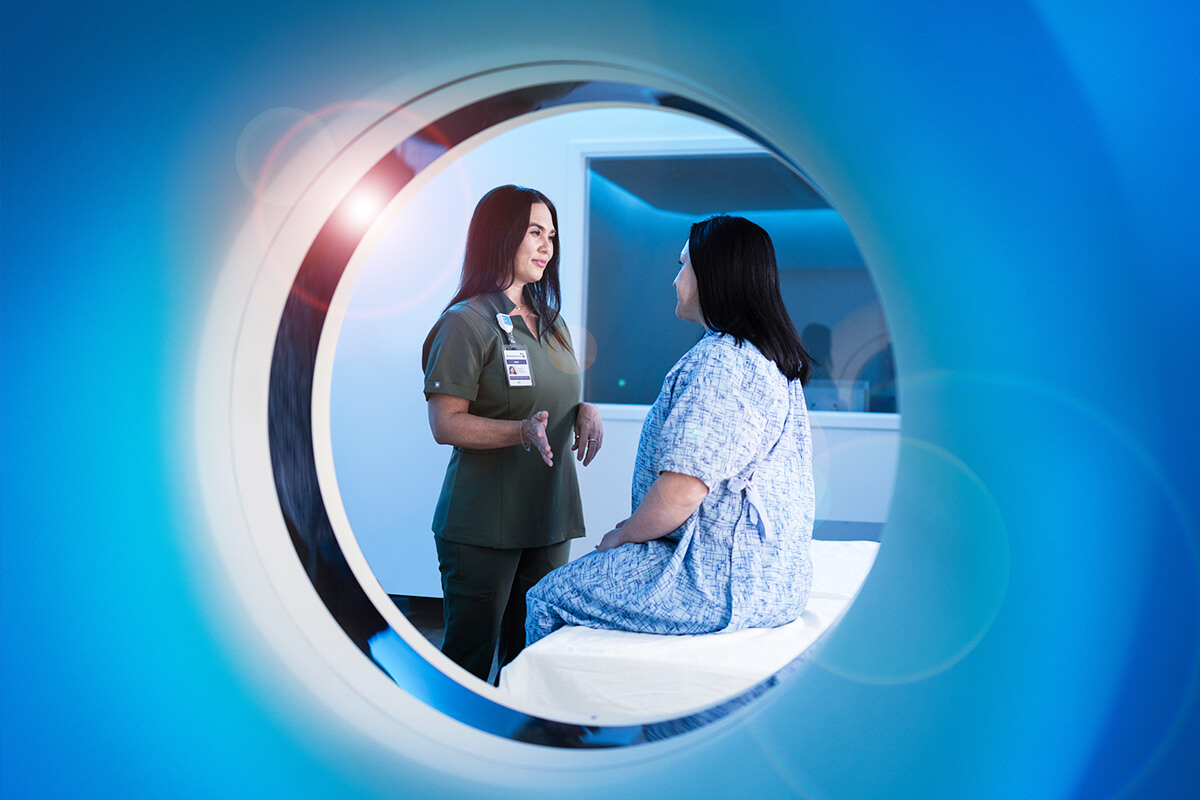
Computed Tomography (CT)
Our CT tests allow your doctor to see a detailed 3D image of the inside of your body. CT can assist in:
- Detecting strokes, head injuries, herniated discs, and abscesses
- Locating fractures
- Determining the extent of bone and soft tissue damage in trauma patients
- Diagnosing changes in various organs
- Examining the heart and coronary vessels
- Diagnosing lung and intestinal cancer earlier
With this non-invasive exam, patients lie on a bed under a circular shaped scanner. The exam typically lasts between 15 and 30 minutes. Our CT technologists will keep you informed throughout the entire process.
Magnetic Resonance Imaging (MRI)
Similar to a CT scan in its design, our magnetic resonance imaging (MRI) creates detailed images of body tissues and is useful for detecting certain diseases that CT scans can't.
With this non-invasive exam, patients lie on a bed under a circular shaped scanner. The open design of our advanced MRI machine is more comfortable, especially for larger patients, and less likely to cause claustrophobia.
A typical MRI scan takes up to one hour. The technologist will ensure you are comfortable in the machine and will be able to hear and talk with you from an adjoining room as your exam is happening.
Mammography and 3D tomosynthesis
Mammograms provide detailed images of breast tissue for detecting breast cancer. Considering that one in eight women will be diagnosed with breast cancer in her lifetime, mammograms are essential for detecting breast cancer early when it is most easily cured.
A mammogram generally takes only 10–15 minutes. The technologist positions the patient in front of a machine that takes images from different angles. The technologist also compresses the breast with a soft, contoured paddle to obtain optimal image quality.
Our imaging centers offer 2D mammograms as well as 3D mammograms (also known as tomosynthesis), which enable a more detailed view and are especially useful for women with dense breast tissue.
Following your exam, a radiologist carefully examines the images for abnormalities and shares the results with your physician.
For your convenience, we offer online scheduling.
Nuclear medicine
With nuclear medicine, a safe radioactive substance (called a radioactive tracer) is introduced into your body either orally or through an injection. This substance is designed to illuminate areas of the body that are then captured by an external camera or scanner. By essentially “lighting up” tissues, organs, or bones, nuclear medicine allows easier and better detection of abnormalities and shows how organs are functioning.
Some common uses include:
- Finding areas of low blood flow through the heart and damaged heart muscle.
- Evaluating the function of the gallbladder and bile ducts.
- Identifying abnormalities within the bones or joints.
- Detecting some forms of cancer
Nuclear medicine exams can take as little as 30 minutes or up to an hour or more.
Positron Emission Tomography (PET/CT)
Positron emission tomography (PET) and computed tomography (CT) are used for cancer diagnosis, treatment planning, and assessment of how well treatments are working. This sophisticated imaging equipment is essential in the treatment and diagnosis of virtually every cancer patient we care for at the Diana J. White Cancer Institute.
A CT scan produces sharp images of cross-sections of your organs and other areas of the body to identify lesions, tumors, and metastases. PET scans focus more on how your organs and internal systems behave rather than on how they look.
Ultrasound
Ultrasound is used for everything from taking a first peek at a developing baby in the womb to determining the risk of vascular disease. By emitting high-frequency sound waves into the body, physicians can translate the echoes that bounce off body tissues and organs into visual images that provide valuable medical information. Ultrasound can also be used to look for gallstones, liver damage, and kidney dysfunction.
X-ray
X-rays are the most commonly used imaging tool, and helpful for diagnosing common bone injuries and diseases, as well as common conditions of the lungs and chest. They are fast and easy, especially in emergency diagnosis and treatment situations. Walk-in X-ray services are available 7 days per week at the main hospital and Monday through Saturday at the Health Pavilion.